OUR BLOG
Valued discussions with our team from wherever you are in your day
FEATURED
EQUINE VETERINARY MEDICAL MANIPULATION
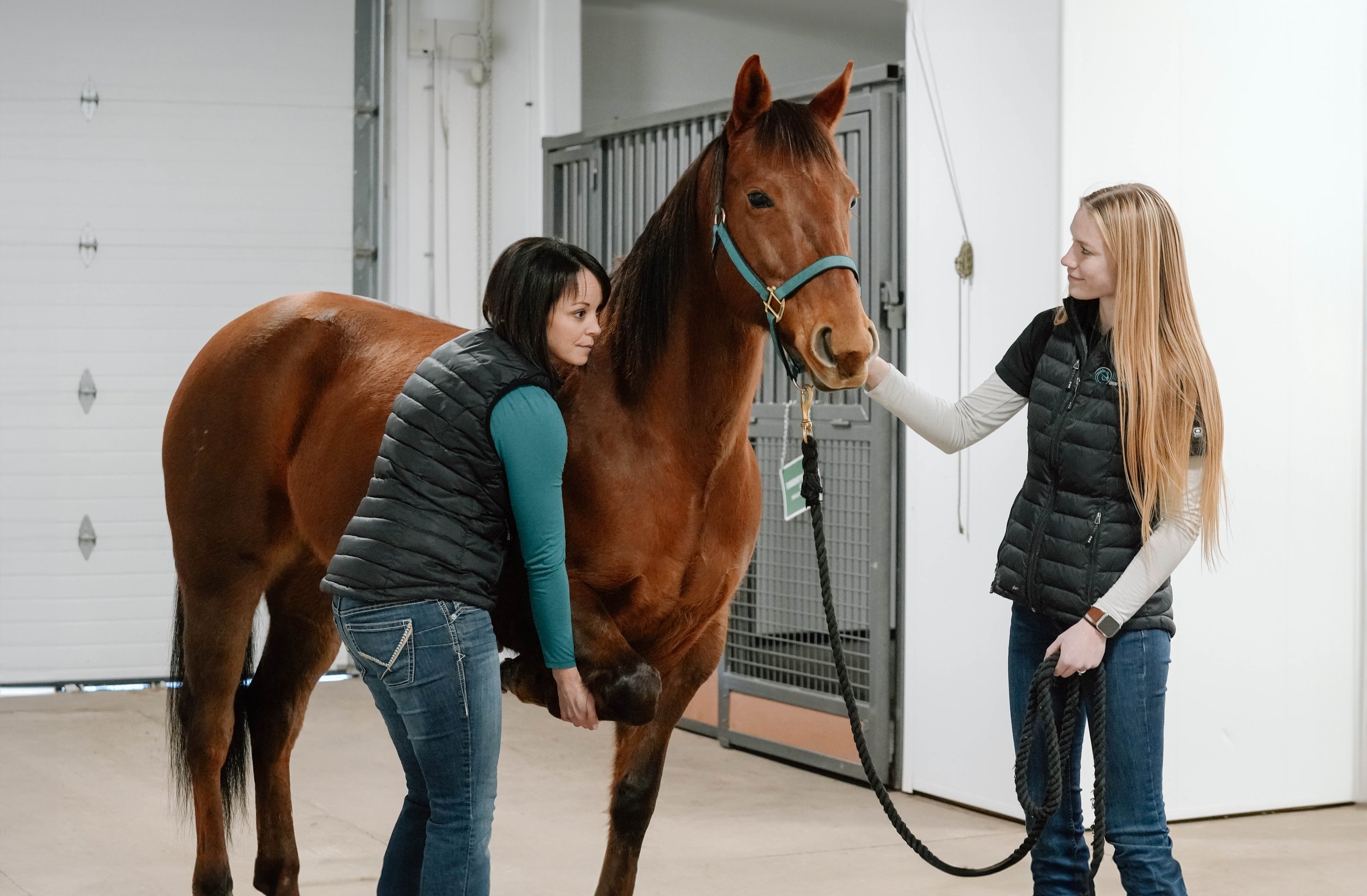
We are excited to announce that Dr. Katy White is now offering equine veterinary medical manipulation (EVMM)!
This manual therapy, similar to chiropractic work or spinal manipulation therapy, includes a full-body assessment of the horse and incorporates spinal manipulation, fascial work, trigger-point therapy, acupressure, and massage. Dr. White completed a certification program with over 150 hours of training from the Chi University in Ocala, Florida that is specifically for veterinarians with a focus on performance horse medicine and rehabilitation.
Misty - Enucleation surgery
An enucleation is the surgical removal of a horse's eye. There are many indications for which this surgery would be performed, including trauma, neoplasia (cancer), extensive infection, or any condition causing pain in a blind eye. In Misty's case, the procedure was recommended due to an acute worsening of uveitis and ulceration along with equine recurrent uveitis flare-ups that had been occurring over the last four years.
Junior - Sarcoid removal
Sarcoids are the most common tumour that occurs in horses. They are locally invasive, and difficult to deal with because recurrence is common even with aggressive therapy. One study showed that 14% of sarcoids occur exclusively in the periocular region (near the eye), and these tumours can be particularly tricky to deal with as it is difficult to get good margins to remove all tumour cells during surgical excision.
Sugar - Skin Grafting
Sugar sustained a major laceration to the front of her carpus during the big snowstorm we had in September 2014. Even though her owner found it the day it happened, there was already a large amount of swelling present as well as a large amount of dirt and contamination in the wound. Initial treatment included intravenous regional limb perfusions with antibiotics, intravenous antibiotics, and bandaging. Because of the large amount of motion present on the front of the carpus, we ultimately decided to use pinch grafts in this wound. Pinch grafts are small 3mm discs of skin, harvested by removing an elevated cone of skin, that are implanted into small slits in the granulation tissue.
Dan - Third Eyelid Removal
In horses, the third eyelid is prone to developing squamous cell carcinoma. Squamous cell carcinoma is the second most common tumour in horses, and it is the most common tumour in the equine eye. It develops most commonly on areas lacking pigmentation, poorly haired regions, and skin near mucocutaneous junctions. It can be quite an aggressive tumour, spreading to nearby tissues and local lymph nodes. In the third eyelid, it often initially appears as a reddened area, then becoming raised and in some cases developing a wart-like appearance. In most other areas, recurrence is extremely common unless surgical excision is combined with another treatment such as chemotherapy or cryotherapy. Fortunately, the third eyelid can be removed in its entirety, and a success rate of 90% has been reported with removal alone.
Stone - Septic Tarsal Sheath
Stone came in to Burwash Equine unable to bear any weight at all on his right hind limb. He had sustained a wound on his hock that had been healing well, but had never been lame prior to the morning he arrived. By palpating the hock, doing an ultrasound exam, taking x-rays, and taking a sample of the fluid from the tarsal sheath, we diagnosed an infection of the tarsal sheath
Luke - Puncture Wound from a Nail in the Foot
When Luke was referred to Burwash Equine by his regular veterinarian, he was unable to bear any weight on his left front limb. His veterinarian had diagnosed a puncture wound to the sole that had likely occurred several days previously. The following x-ray shows a probe placed in the puncture wound to demonstrate which structures in the foot may have been involved. Although the probe doesn’t extend the entire path of the wound, from this x-ray and taking a sample of fluid from the digital tendon sheath, we suspected infection of both the navicular bursa and the tendon sheath.
Roy - Carpal Laceration
Roy sliced the skin off the front of his knee slipping on a rubber mat over the Easter weekend. We injected saline into both his radiocarpal joint and his middle carpal joint to make sure they weren't involved in the wound, and then were able to close the wound with sutures.
TOPIC SUBMISSIONS
Want to learn more about a specific topic? Email your ideas to our team: office@burwashequine.ca
Archive
-
Breeding
- Mar 6, 2018 Placentitis - a reason for monitoring your pregnant mare
- Mar 6, 2017 Breeding Your Mare
- Mar 20, 2015 Breeding Your Mare: A behind-the-scenes look at the science of mare reproduction
- Nov 19, 2014 Fall Seminar 2014: An Introduction to the World of Reproduction
- Nov 10, 2014 Stallion Semen Freezing
- Oct 16, 2014 Pregnant Mare Management
- Apr 24, 2014 Breeding Your Mare
-
Dentistry
- Jan 25, 2020 EOTRH: a dental disease in the elderly equine
- Nov 25, 2019 Proactive Winter Horse/Donkey/Mule Care
- Jan 22, 2017 What happens to wild horses that don't get dental care?
- Apr 25, 2014 Equine Dentistry: Why Equine Veterinarians are Uniquely Qualified
- Apr 25, 2014 A Guide To Equine Dental Care
-
Deworming
- Nov 25, 2019 Proactive Winter Horse/Donkey/Mule Care
- Feb 13, 2015 Nasty Little Parasites - An Update on Deworming
- Oct 16, 2014 Parasite Control Recommendations
-
Emergencies
- Aug 29, 2024 Equine Emergencies
- Sep 1, 2021 Getting Back to Better by Dr. Crystal Lee
- May 8, 2016 First Aid Seminar - Part 2
- May 8, 2016 First Aid Seminar - Part 1
- Apr 15, 2016 First Aid Seminar Slides
- Oct 5, 2014 Brio - Heel Bulb Laceration
- Apr 27, 2014 Stone - Septic Tarsal Sheath
- Apr 27, 2014 Luke - Puncture Wound from a Nail in the Foot
- Apr 26, 2014 Roy - Carpal Laceration
- Apr 23, 2014 Be Prepared for an Equine Health Emergency
-
Foals
- Apr 17, 2020 Fall Seminar 2019 - The Events of Normal Foaling
- Mar 6, 2018 Placentitis - a reason for monitoring your pregnant mare
- May 24, 2016 My Newborn Foal
- Apr 16, 2016 When Foaling Is Imminent
- Apr 6, 2016 When is my mare going to foal?
-
Lameness
- Mar 14, 2023 Steve with no sole
- Nov 24, 2022 Regenerative Medicine and Orthobiologics
- Jun 1, 2022 Osteoarthritis by Dr. Katy White
- Jul 1, 2020 Recognizing and Managing the Club Foot in Horses
- Jul 1, 2018 Defying Age
- Jan 23, 2016 Fall Seminar 2015 - Update on the Lameness Locator
- Sep 13, 2015 Focus on Lameness - Imaging
- Sep 13, 2015 Focus on Lameness - Lameness Locator Demonstration
- Sep 13, 2015 Focus on Lameness - The Lameness Locator
- Sep 13, 2015 Focus on Lameness - General Lameness Exam
- Apr 11, 2015 Equine Lameness Evaluation
- Mar 25, 2015 Wireless inertial sensor based objective lameness evaluation - seminar slides
-
Medicine
- Dec 27, 2024 Why Your Horse Needs Vitamin E
- Aug 29, 2024 Equine Emergencies
- Nov 24, 2022 Regenerative Medicine and Orthobiologics
- Jun 1, 2022 Osteoarthritis by Dr. Katy White
- Feb 1, 2022 Respiratory Disease Round-Up
- Nov 13, 2019 Strangles
- Dec 20, 2018 Understanding PPID
- Jul 1, 2018 Defying Age
- Feb 1, 2016 Fall Seminar 2015 - Equine Infectious Anemia
- Sep 16, 2015 Flor - Pituitary Pars Intermedia Dysfunction (PPID)
- Nov 21, 2014 Fall Seminar 2014: Pigeon Fever Updates
- Nov 4, 2014 Fall Seminar 2014: Common Conditions of the Equine Eye
-
News
- Nov 24, 2022 Regenerative Medicine and Orthobiologics
-
Podiatry
- Mar 14, 2023 Steve with no sole
- Jul 1, 2020 Recognizing and Managing the Club Foot in Horses
- Nov 12, 2016 Fall Seminar 2016 - A Snapshot of the Horse's Foot - Equine Podiatry
-
Prepurchase Exams
- Nov 5, 2016 Fall Seminar 2016 - An Overview of Prepurchase Exams
- Apr 23, 2014 The pre-purchase exam: A wise investment
-
Surgery
- May 14, 2025 Nodin - Incisor Wiring
- Sep 1, 2021 Getting Back to Better by Dr. Crystal Lee
- Sep 26, 2018 Colic Surgery
- Oct 14, 2015 Misty - Enucleation surgery
- Sep 21, 2015 Junior - Sarcoid removal
- Sep 8, 2015 Sugar - Skin Grafting
- Aug 31, 2015 Dan - Third Eyelid Removal
-
Vaccines
- Nov 25, 2019 Proactive Winter Horse/Donkey/Mule Care
- Nov 13, 2019 Strangles
- Nov 5, 2016 Fall Seminar 2016 - An Update on West Nile Virus and Rabies in Alberta
- Feb 9, 2016 Fall Seminar 2015 - Vaccines
- Apr 30, 2015 Vaccination FAQ - What are common side effects of vaccination? What can I expect after my horse is vaccinated?
- Apr 28, 2015 Why We Vaccinate - Equine Herpesvirus (“Rhinopneumonitis”)
- Apr 24, 2015 Vaccination FAQ - Why are horses vaccinated for tetanus yearly, whereas humans are boostered every 5-10 years?
- Apr 23, 2015 Vaccination FAQ - Why is the Strangles vaccine intranasal? Isn’t there an intramuscular vaccine available?
- Apr 22, 2015 Rabies in Alberta—Should We Be Vaccinating Horses?
- Apr 21, 2015 Why We Vaccinate - Equine Influenza
- Apr 19, 2015 Vaccination FAQ - Potomac Horse Fever?
- Apr 18, 2015 Vaccination FAQ - Is it better to give multiple vaccines on one date, or split them into different visits?
- Apr 17, 2015 Vaccination FAQ - My horse needs his hocks injected, and while you are here, could we vaccinate him as well?
- Apr 16, 2015 Why We Vaccinate - Strangles
- Apr 14, 2015 Vaccination FAQ - Is it okay to ride my horse immediately before or after he or she is vaccinated?
- Apr 13, 2015 Why We Vaccinate - West Nile Virus
- Apr 12, 2015 Vaccination FAQ - Can I have my horse vaccinated if (s)he has a mild “cold?”
- Apr 11, 2015 Why We Vaccinate - Eastern & Western Equine Encephalitis (“Sleeping Sickness”)
- Apr 11, 2015 Vaccination FAQ - My horse is vaccinated—why did it still get a cold?
- Apr 9, 2015 Why We Vaccinate - Tetanus in Horses
- Apr 7, 2015 Vaccination FAQ - My horse doesn’t go anywhere—does (s)he still need to be vaccinated?
- Apr 6, 2015 What's in my vaccine?
- Apr 14, 2014 Vaccination Protocols
-
Wellness
- Jun 1, 2022 Osteoarthritis by Dr. Katy White
- Feb 1, 2022 Respiratory Disease Round-Up
- Jan 18, 2022 Baled It! with Dr. Lauren Friedl
- Sep 1, 2021 Getting Back to Better by Dr. Crystal Lee
- Nov 25, 2019 Proactive Winter Horse/Donkey/Mule Care
- Dec 20, 2018 Understanding PPID
- Jul 1, 2018 Defying Age
- Feb 14, 2018 Sorting Through Supplements - how to tell if the supplement is worth buying
- Jan 8, 2017 Fall Seminar 2016 - Senior Horses - A Focus on the Care and Quality of Life of Older Horses
- Dec 8, 2016 Saying Goodbye: a discussion about euthanasia
- Jul 1, 2015 The Equine Eye by Dr. Kirby Penttila
- Apr 11, 2015 Equine Lameness Evaluation
- Oct 16, 2014 Forage Alternatives


